

The above statements are generalizations based on scientific analyses of large population data sets, such as survivors exposed to radiation from the atomic bomb. body region - Some organs are more radiosensitive than others.patient’s sex - Women are at a somewhat higher lifetime risk than men for developing radiation-associated cancer after receiving the same exposures at the same ages.patient’s age - The lifetime risk of cancer is larger for a patient who receives X-rays at a younger age than for one who receives them at an older age.

radiation dose - The lifetime risk of cancer increases the larger the dose and the more X-ray exams a patient undergoes.The risk of developing cancer from medical imaging radiation exposure is generally very small, and it depends on:
Mri vs xray skin#
For example, the typical use of a CT scanner or conventional radiography equipment should not result in tissue effects, but the dose to the skin from some long, complex interventional fluoroscopy procedures might, in some circumstances, be high enough to result in such effects.Īnother risk of X-ray imaging is possible reactions associated with an intravenously injected contrast agent, or “dye”, that is sometimes used to improve visualization.

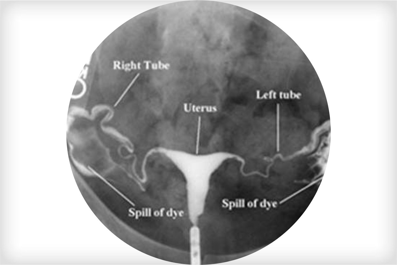
CT - many X-ray images are recorded as the detector moves around the patient's body.Fluoroscopy can result in relatively high radiation doses, especially for complex interventional procedures (such as placing stents or other devices inside the body) which require fluoroscopy be administered for a long period of time. Fluoroscopy - a continuous X-ray image is displayed on a monitor, allowing for real-time monitoring of a procedure or passage of a contrast agent (“dye”) through the body.Mammography is a special type of radiography to image the internal structures of breasts. Radiography - a single image is recorded for later evaluation.Ionizing radiation is a form of radiation that has enough energy to potentially cause damage to DNA and may elevate a person’s lifetime risk of developing cancer.ĬT, radiography, and fluoroscopy all work on the same basic principle: an X-ray beam is passed through the body where a portion of the X-rays are either absorbed or scattered by the internal structures, and the remaining X-ray pattern is transmitted to a detector (e.g., film or a computer screen) for recording or further processing by a computer. Computed tomography (CT), fluoroscopy, and radiography ("conventional X-ray" including mammography) all use ionizing radiation to generate images of the body. There are many types - or modalities - of medical imaging procedures, each of which uses different technologies and techniques. Medical imaging has led to improvements in the diagnosis and treatment of numerous medical conditions in children and adults. Medical device requirements for manufacturers of x-ray imaging devices.Electronic Product Radiation Control (EPRC) requirements for manufacturers and assemblers.Regulations and guidelines pertaining to imaging facilities and personnel.Information for the referring physician.Principles of radiation protection: justification and optimization.Questions to ask your health care provider.


 0 kommentar(er)
